Student Research Project
Capacitive Tools for Human Cell Analysis
June 2022, Hamburg
Project Objectives

Advancing our biotechnological capabilities is crucial for diagnostic and therapeutic tools. Capacitive microwave sensors unlock early-stage monitoring to a wide branch of currently overseen diseases by providing insights into the clockwork of human cells.
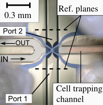
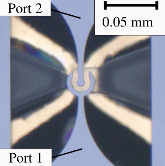
What branches of science have the potential to break these boundaries? Researchers from Hamburg University of Technology cooperated for our assistance in the development of fluidic microchips precisely measuring the broadband permittivity of single cells. Our technology features a three-way mechanical trap to capture and position single cells while reducing the parasitic contribution of the surrounding medium. This novel approach has the potential of saving real lifes.
Brief Introduction

The capacitive microwave sensor and its guard electrode are trimmed for maximized information gains on specific cells and both minimized design and impact on biological environment. Key technology for those achievements is self-designed, trailed and optimized.
I. How do capacitive microwave sensors work?
Built upon monitoring an electric fields capacitance, this extended technology meassures electric fields changes inbetween multiple conductive plates – an indispensable bit of information to review the materials dielectric properties.
Further, including guard electrodes around the capacitor plates edges minimizes the fringing fields distortion on highly precise meassurements. Connected to a high-impedance amplifier, its total gap in potential to the sample is meassured and the voltage amplified. This increases accuracy by orders of magnitude.
II. What precise cell parameters are being captured?
Both size and shape of the cell enable researchers determining cell typus, stage of development and internal anomalies. Permitivity unveils the cells and itts membranes compositionl – the foundation for assessing its overall health. Other parameters include intracellular and extracellular ion concentrations (highly relevant information regarding cell functionality and metabolism), mechanical properties (stiffness, elasticity, and viscosity), and fluorescence and marker expression (useful for quantifying and tracking cellular components, protein expressions, and gene activity).
Optimizing Cutting-Edge Sensors


Various patterns of established hardware have been optimized to provide more accurate, precise and reliable data.
Sensor Architecture: Miniaturization of vital hardware got achieved by utilizing commercial processes involving gold-plated glass wafers carrying electrodes. Efficient design patterns made up by layers of photoresist (SU8) host the microfluidic channel, enabling single-cell measurements and ensuring proper contact between cells and the sensing erea while keeping their free movement elsewhere running.
Multi-Layer Design: The microchip involves stacked metal structures, microfluidic channels and ground electrodes, directly connected to through coplanar waveguides (CPW). This level of performance is engineered using early-stage tools like Ansys HFSS. Gold-plated glass wafers ensure consistency in reliability.
Verification Setup: The research has devised the method of assessing reliability by introducing a chip holder, attached tubing (enabling controlled fluid flow) and measurement probes (capturing the cells electrical properties by establishing electrical connections).
Impedance Considerations: Exceptionally thin metal layers cloud characteristic impedances. Therefore, calibration methods are crucial to not fall for the illusion of possible cross dependencies between capacitance and conductance.
The success of the self-designed and -coded hardware speaks for itself: It provides scientists with both accurate and reliable data sets over extensive experimental periods. This highly optimized technology effectively differentiates between various fluids and cells. Extracted permittivity align with validated data, indicating accuracy and effectiveness.
Additionally, single-cell measurements have been conducted on both living and dead cells. The sophisticated recognition and contextual classification of numerous cellular properties from the perspective of a millimeter-sized chip represents a great scientific accomplishment.
Related Researchers


